Wednesday, September 17, 2008

Common Rashes With Blisters


Do you know what's causing those blisters? Should you see a doctor or can you take care of it on your own? Take a look at some common rashes with blisters and see if you can find yours.

Chicken Pox:

Chicken pox is a highly contagious viral infection caused by the varicella virus. The word chickenpox comes from the Old English word "gican" meaning "to itch" or from the Old French word "chiche-pois" for chickpea, a description of the size of the lesion.

Who Gets Chicken Pox
Chickenpox is a disease of childhood - 90% of cases occur in children aged 14 years and younger. Before widespread vaccination, the incidence of chicken pox in the United States approached the annual birth rate, averaging between 3.1 and 3.8 million cases per year. Chicken pox can occur at any time, but occurs most often in March, April, and May in temperate climates.

Varicella Virus
The varicella virus is an enveloped, double-stranded DNA virus. It attaches to the wall of the cell it invades, and then enters the cell. The virus uncoats and is transported to the nucleus where the viral DNA replicates creating new virions that are eventually released from the cell to infect other cells.

Acquiring Chicken Pox
Chicken pox is acquired by direct contact with infected blister fluid or by inhalation of respiratory droplets. When a person with chicken pox coughs or sneezes, they expel tiny droplets that carry the varicella virus. A person who has never been exposed to chicken pox inhales these droplets and the virus enters the lungs, and then is carried through the bloodstream to the skin where it causes a rash. While the virus is in the bloodstream (before the rash begins) it causes typical viral symptoms like fever, fatigue, joint pains, headache, and swollen glands. These symptoms usually resolve by the time the rash develops. The incubation period of chicken pox averages 14 days with a range of 9 to 21 days.

Appearance of Chicken Pox
The chicken pox rash begins on the trunk and spreads to the face and extremities. The chicken pox lesion starts as a 2-4 mm red papule which develops an irregular outline (rose petal). A thin-walled, clear vesicle (dew drop) develops on top of the area of redness. This "dew drop on a rose petal" lesion is very characteristic for chicken pox. After about 8-12 hours the fluid in the vesicle gets cloudy and the vesicle breaks leaving a crust. The fluid is highly contagious, but once the lesion crusts over, it is not considered contagious. The crust usually falls off after 7 days sometimes leaving a craterlike scar. Although one lesion goes through this complete cycle in about 7 days, another hallmark of chicken pox is the fact that new lesions crop up every day for several days. Therefore, it may take about a week until new lesions stop appearing and existing lesions crust over. Children are not sent back to school until all lesions have crusted over.

Poison Ivy:


Poison ivy is known in medical terms as Rhus Dermatitis which is a type of contact dermatitis. As the name implies, a contact dermatitis is an irritation of the skin caused by contact with a specific irritant. In the case of poison ivy, the irritant is called urushiol which is a resin found in the plants in the Anacardiaceae family and the Rhus genus. Plants included in this classification are poison ivy, poison oak, and poison sumac. Also included are the cashew nut tree, mango tree, Japanese lacquer tree, and marking nut tree.

Poison Ivy
The appearance of poison ivy, oak, and sumac varies by regions and season. Poison ivy leaves are most likely to be in groups of three and notched, although they can be smooth edged. Poison ivy is usually found east of the Rocky Mountains growing as vines or shrubs.

Poison Oak
Poison oak leaves come in groups of three, five, or seven. They are smaller than poison ivy leaves and have smooth, rounded edges. Poison oak is usually found west of the Rocky Mountains as a small bushy plant or climbing vine.

Poison Sumac
Poison sumac has seven to thirteen leaves on one stem angled upward. They are smooth edged, oval and about 10 cm long. Poison sumac is found in boggy areas in the south.

Interesting Facts About Poison Ivy
In the United States poison ivy, poison oak, and poison sumac cause more cases of contact dermatitis than any other agents combined. Rhus dermatitis accounts for 10% of the US Department of Agriculture and Forestry Services lost time injuries. Twenty-five million to 40 million Americans require medical attention after being exposed to one of these plants.

Poison Ivy occurs from contact with the leaf or internal parts of the stem or root of the plant. Eight to 48 hours after exposure to urushiol the characteristic rash appears. This rash is typically red, contains blisters, and is in a linear or circular pattern. Urushiol can be found under fingernails, on clothing, and on tools unless it is deliberately removed. The resin itself can be active and cause a new rash for up to 3 weeks after exposure. Urushiol is not found in blister fluid and not responsible for spreading the rash. If untreated, the rash usually resolves in 3 weeks.

Treatment of Poison Ivy
The most common sites on the body for poison ivy are exposed areas on the arms, legs, and face. The intensity of the rash varies depending on the sensitivity of the person, and the amount and extent of exposure.

  • Washing the skin with soap and water inactivates and removes the resin. Washing is most effective if it is done within 15 minutes of exposure.
  • Cold, wet compresses are effective in the blistering stage. They should be used for 15 to 30 minutes several times a day for the first 3 days.
  • Steroid creams or ointments are helpful to reduce redness and itching. Hydrocortisone can be used on the face, but is usually not strong enough for more than mild cases on the arms or legs. Typically, a prescription strength steroid is needed for these areas.
  • Oral steroids are used for severe cases of poison ivy but must be used for at least a week.
  • Short, cool tub baths with colloidal oatmeal (Aveeno) can be soothing and help control inflammation.
  • Calamine lotion helps control itching but used too long can cause excessive drying of the skin and more inflammation.
  • Antihistamines help reduce itching and the older types such as diphenhydramine (Benadryl) help encourage sleep.
  • Any exposure to the eyes or eyelids or the development of a honey-colored crust should be evaluated by a health care provider.

Prevention of Poison Ivy

  • Desensitization is not effective either by chewing leafs or having commercially prepared extracts injected.
  • The most effective prevention is using a barrier to protect the skin. Clothing serves as an effective barrier but since the urushiol remains on the clothing, it must be removed carefully and laundered without contacting the skin.
  • Urushiol can penetrate latex gloves but not rubber gloves.
  • A lotion containing 5% quaternium-18 bentonite (IvyBlock) can be applied to the skin and provides a barrier for 4 to 8 hours. It must be washed off and reapplied for continued exposure.

The First Genital Herpes Outbreak:
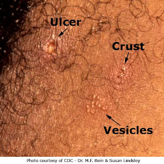

Most people who are infected with the herpes simplex virus do not have symptoms. Of those who do develop symptoms, the first outbreak of genital herpes is worse than recurrences. The first outbreak is also associated with general symptoms aside from the rash. Women are at risk

of having a herpes infection that does not cause the usual symptoms.

Genital Herpes and Herpes Simplex Viruses
In the past, genital herpes was caused mainly by the herpes simplex virus type 2 (HSV-2). But now, new genital herpes infections are caused equally by the herpes simplex virus type 1 (HSV-1) and the herpes simplex virus type 2 (HSV-2). The majority of people who are going to get a primary outbreak will do so between 3 days to 2 weeks after exposure.

The First Genital Herpes Outbreak
The rash of herpes is a cluster of vesicles on a red base. In moist areas like the vagina, herpes may cause ulcerations instead of blisters. In women, the first outbreak of genital herpes can occur on the vulva, cervix, vagina, urethra, anus, buttocks, or thighs. Men usually get an outbreak on the tip of the penis or the shaft, but rarely around the base. Men who have sex with men may also get blisters in or around the anus. The rash in men is usually mild -- only 6 to 10 blisters. The blisters in men and women are painful and contain a large number of viral particles; therefore, they are very contagious.

Other Symptoms With the First Genital Herpes Outbreak
Seventy-nine percent of people get general symptoms with the first outbreak that usually resolve within a week. Some common general symptoms include:

  • Fever to 102 degrees
  • Headache
  • Muscle aches
  • Fatigue
  • Swollen lymph nodes

Women and the First Genital Herpes Outbreak
Women are four times more likely to be infected with HSV-2 than men. For some reason, women have more severe disease and more complications during the first infection than men. If a woman gets a herpes outbreak on the cervix or vagina and not externally, she may develop vaginal discharge, pelvic pain, or burning with urination. With the first outbreak, some women may get a second round of blisters or ulcers in the second week.

How Long the First Genital Herpes Outbreak Lasts
The first herpes infection usually lasts for 2 to 3 weeks, but skin pain can last for 1 to 6 weeks. The blisters dry out and crust over. When the crusts fall off, the area is usually not contagious anymore. There is evidence that some people have low levels of virus present even when they do not have symptoms.

Symptoms Checker

Elbow Pain:

We're sorry you have elbow pain!

Please take a few minutes to answer some questions and this guide will provide some of the more likely causes and provide some helpful information. The guide is not intended to replace a face-to-face evaluation with your doctor.

The diagnoses provided are among the most common that could explain your symptoms, but the list is not exhaustive and there are many other possibilities. In addition, more than one condition may be present at the same time. For example, a person with rheumatoid arthritis could also have ulnar neuropathy because swelling in the elbow compresses the nearby nerve.

Do you have severe elbow pain as well as any of the following symptoms:

  • fever
  • redness
  • marked swelling
  • inability to use the joint
  • or recent significant trauma (a fall, car accident, etc.)?

Gout:

We're sorry to hear you have gout (or may have gout).

The goal of this guide is to provide information while awaiting evaluation with your doctor or additional information after you have seen him or her. Please keep in mind that this guide is not intended to replace a face-to-face evaluation with your doctor.

First, some background information about this guide and about the condition itself:

Gout is a condition in which one or more joints become inflamed when crystals of urate (also called uric acid) deposit there. Urate is a byproduct of normal bodily functions and is removed from the body by the kidneys.

This guide will ask you a series of questions and depending on your answers, information will be provided and additional questions asked until the conclusion where a series of links for more information will be provided. Or you may choose additional general information about gout.

Hand Pain:

Sorry to hear you have hand pain.

The goal of this guide is to provide information while awaiting evaluation with your doctor, or for additional information after you have seen him or her. Please keep in mind that this guide is not intended to replace a face-to-face evaluation with your doctor.

Hand pain may develop for a number of reasons -- fracture and infection are among the most serious while sprains and strains are among the most common. At the end of this guide, you will see links to more detailed information about the most common causes of hand pain; however, there are rare causes of symptoms that will not be included here and would require more detailed evaluation than this guide can provide.

Certain symptoms suggest a serious cause of hand pain that requires prompt attention. It's important to ask questions about these symptoms first.

Nail Problems:

Sorry to hear you have a problem with your nail(s).

Please keep in mind that this guide is not intended to replace a face-to-face evaluation with your doctor. The goal of this guide is to provide information while awaiting evaluation with your doctor or additional information after you have seen him or her.

Nail problems may develop for a number of reasons -- skin inflammation, an ingrown nail, and psoriasis are among the most common, but an infection is among the most serious. Any problem in the nail that does not improve over time should be evaluated by your physician.

At the end of this guide, you will be offered links to more detailed information about the most common nail problems; however, there are rare causes that will not be included here and would require more detailed evaluation than this guide can provide.

Raynaud's:

Welcome to the Raynaud's Decision Guide.

We're sorry to hear you have Raynaud's!

The goal of this guide is to provide information while awaiting evaluation with your doctor, or additional information after you have seen him or her. Please keep in mind that this guide is not intended to replace a face-to-face evaluation with your doctor. The diagnoses provided are among the most common that could explain your symptoms, but the list is not exhaustive and there are many other possibilities. In addition, more than one condition may be present at the same time. For example, a person with Raynaud's could also have osteoarthritis that is unrelated.

This guide is intended for persons who have Raynaud's -- you may hear it called Raynaud's phenomenon, disease, or syndrome. Whatever the name, the basic problem in this condition is vasospasm -- that is, a blood vessel (the "vaso" part) suddenly constricts (the "spasm" part), usually after exposure to cold. Then, not enough blood can flow to the finger, toe or whatever part is affected. This is an exaggeration of the normal behavior of blood vessels. Tiny nerves tell the artery to open or close, depending on the situation. Usually with cold exposure, blood vessels constrict, or close a bit, to conserve heat, but for people with Raynaud's, that mechanism is overactive. The cause is not known. People with Raynaud's notice intensely cold fingers or toes, numbness, pain, and a color change -- typically from white to blue to red, and, eventually, back to normal, over a number of minutes.

Most people with Raynaud's (up to 90 percent or more) are otherwise healthy and have no other medical conditions. Raynaud's is quite common -- up to ten percent of healthy young women report symptoms suggestive of Raynaud's. The associated conditions, on the other hand, are rather rare, and include lupus, scleroderma and CREST syndrome, among others. There will be an opportunity to learn more about these conditions at the end of this guide.

Now, let's move on to the specifics of your situation.

Besides the Raynaud's, do you have one or more of the following:

  • tight, thickened skin over the fingers, hands, arms and/or face
  • pain in multiple joints that is worse in the morning
  • a chronic (long-standing) rash
  • chronic dry eyes and dry mouth
  • chest pain when you take a deep breath
  • severe reflux disease (also called GERD, a form of heartburn)?
Shoulder Pain:

We're sorry you have shoulder pain!

The goal of this guide is to provide information while awaiting evaluation with your doctor, or for additional information after you have seen him or her. Please keep in mind that this guide is not intended to replace a face-to-face evaluation with your doctor. The diagnoses provided are among the most common that could explain your symptoms, but the list is not exhaustive and there are many other possibilities. In addition, more than one condition may be present at the same time. For example, a person with rheumatoid arthritis could also have tendonitis.

The shoulder is prone to developing pain in part because its anatomy is complex and because it has the largest range of motion of any joint in the body. Some of these muscles, tendons, and bursae are common causes of shoulder pain, even when the joint itself is fine.

Do you have severe shoulder pain with the following symptoms:

  • fever
  • redness
  • marked swelling
  • inability to use the joint
  • recent significant trauma (a fall, car accident, etc.)?
Wrist Pain:

We're sorry you have wrist pain!

The goal of this guide is to provide information while awaiting evaluation with your doctor, or for additional information after you have seen him or her. Please keep in mind that this guide is not intended to replace a face-to-face evaluation with your doctor. The diagnoses provided are among the most common that could explain your symptoms, but the list is not exhaustive and there are many other possibilities. In addition, more than one condition may be present at the same time. For example, a person with rheumatoid arthritis could also have tendonitis.

The wrist is prone to pain because it is frequently overused and has a complicated anatomy, with many structures packed into a small space.

Certain symptoms suggest a serious cause of wrist pain that requires prompt attention. It's important to ask questions about these symptoms first.

Do you have severe wrist pain and any of the following symptoms:

  • fever
  • redness
  • marked swelling
  • inability to use the joint
  • recent significant trauma (a fall, car accident, etc.)?

Monday, September 15, 2008

Breast Cancer Symptoms

Breast cancer is a disorder in which malignant or cancer cells are formed in the breast tissues of women. This is a heterogeneous disease which is found to be different in different women and the symptoms too vary accordingly. Women are advised to have themselves medically examined completely at least once in six months. You can even make yourselves aware of the symptoms of breast cancer and contact the physician immediately if you should notice any at any point of time. Prevention is better than cure; here in this case early detection of the condition will help in faster cure. By knowing the symptoms you can regularly examine yourself for the incidence of breast cancer.
Main Symptoms of Breast Cancer:-
Some of the most common breast cancer symptoms include the following:
In most cases the initial symptom which can be noted would be a lump either on the breast itself or in the armpit. This would be easily noticeable and the patient would be able to find it herself. The lump would feel little thicker than the other tissues of the breast. If this is noticed, whatever be the cause, it is advisable to contact the doctor for an examination.
There might also be a change in shape and size of the breast. Though changes in the size and shape of the breast might also be due to increase or decrease in body weight and shape, this would be more evident and easily noticeable.
Skin dimpling is also a symptom which is noted in many cases.
Another common symptom that can be noticed is inversion of the nipple. The nipple might look as if it is dumped inside. This would really change the shape of the breast itself making it look very odd.
Pain as a symptom, however, cannot be relied upon as it might also be due to some other medical conditions related to breast.
When dermal lymphatics are invaded by the cancer cells, there might be lump formation over the breast also causing inflammation and burning of the nipples. Pain, redness and swelling might also accompany the condition with a change in the skin texture. This might resemble the texture of orange and this condition is referred to us as ‘peau d’orange’.
Paget’s disease of the breast is another stage in which there might be severe itching, burning and pain. Though these are the common symptoms, these need not necessarily be the only ones. These are the most common breast cancer symptoms. They can usually be detected in the early stage itself but if let unnoticed, breast cancer will either lead to mastectomy (removal of the affected breast/s) or death. It is best to check with the physician if you should notice any change in the normal size and shape of the breast.

Summary of most common symptoms:

  • Tender nipples
  • Lumps over the breasts
  • Swelling over the breast
  • Change in appearance of the breast
  • Pain in the breast
  • Nipple discharge in only one of the breasts

Find information on Breast Cancer Treatment here.

Thursday, September 11, 2008

Breast Cancer Surgeries


Healthy Breast Tissue

A mastectomy is the surgical removal of a breast.

The vast majority of mastectomies are performed on women as a treatment for breast cancer, although men may develop breast cancer and require the procedure, too. In some cases, a lumpectomy may be another surgical treatment option for these patients. Those with more extensive breast cancer may require a bilateral mastectomy, which is the removal of some or all of the tissue in both breasts.

Some men with a condition called gynecomastia, in which the breast tissue becomes overdeveloped, opt for a mastectomy for cosmetic purposes.


There are several types of mastectomies. The decision of what type of mastectomy should be done should be made with the assistance of the surgeon performing the mastectomy, the oncologist and the plastic surgeon performing the reconstruction. Some techniques may not be able to be considered, depending upon the location and severity of the cancer. The emotional needs of the patient must also be weighed. 

The decision to have breast reconstruction surgery should be made before the mastectomy surgery is done. The reconstruction may be able to be done right after the mastectomy or at a later date. 

The surgery is done under general anesthesia and typically lasts 2 to 3 hours, although it may take longer if a procedure to remove the lymph nodes is planned or reconstructive surgery is being performed immediately after the mastectomy.

Breast Lump Before Surgery

What Is a Lumpectomy..

A lumpectomy is a surgical procedure to remove a small area of breast tissue that is cancerous or suspected to be cancerous. The lumpectomy procedure is known by several names, including breast conservation surgery, excisional biopsy and partial mastectomy. 

Lumpectomy surgery is typically performed by a surgical oncologist, a physician who specializes in the treatment of cancer with surgical interventions. The surgery can be performed on an inpatient or outpatient basis. If general anesthesia is used, the patient typically remains in the hospital overnight. If local anesthesia is used, the patient may go home the same day as the surgery. 

This surgery is typically reserved for women who have a single, relatively small area of tissue that must be removed. Women with a large mass may need to have significantly more breast tissue removed, which often requires a more aggressive approach such as a mastectomy. 

While a lumpectomy is less disfiguring than a mastectomy and typically does not require reconstructive plastic surgery afterward, a lumpectomy can cause significant changes in the appearance of the breast if a large amount of tissue is taken. In addition to the scar or scars left by surgery, there may be a visible change in skin texture over the site. There may also be an obvious lack of tissue in the area, even after the skin heals.

Lumpectomy Surgery

Lumpectomy Breast Surgery..

After the skin is prepared for the procedure and anesthesia is given, the surgery begins with an incision over the site of the tissue to be removed. Once the skin is opened and the tissue that is to be removed identified, the surgeon will inspect the mass to determine what type of tissue it is made of. 

In some cases, the mass may actually be a cyst, a small fluid-filled sac. If it is a cyst, the fluid will be aspirated until the cyst is emptied of fluid. The fluid will be saved for an analysis after the surgery. 

In most cases, the lump of tissue is not a cyst and will be removed from the breast. In addition to the suspected cancerous tissue, the area around the mass will also be removed, an area called a margin. All of the tissue is saved for analysis later by a pathologist. The surgeon will also inspect the area to see if any additional structures of the body are affected by the lump, such as the muscle underlying the breast. 

After the removal of the tissue, the surgeon will inspect the remaining breast tissue for any signs of cancer that may have spread outside of the lump that was removed. If there is no indication of further problematic tissue, the incision can be closed with sutures and the surgery is finished. 

For some patients, the surgeon will also remove lymph nodes or take samples of lymph nodes for testing. This requires a separate incision under the arm. The surgeon may sample several lymph nodes or remove as many as 15 or 20 nodes in their entirety, depending on the patient’s needs. This is done to determine if cancer has spread from the initial mass in the breast, if it is cancerous. 

The breast will be covered with a sterile bandage during the procedure, as will the underarm incision if lymph nodes are removed.

After Lumpectomy

After Lumpectomy Surgery

After the surgery is completed and the patient has fully awakened from anesthesia, the recovery from surgery truly begins. If lymph nodes were removed, a drain is placed in the underarm area to remove excess fluid. The drain will be under a sterile bandage, and can be easily removed after surgery when the drainage is minimal. 

Dissolvable sutures may be slowly absorbed into the body over time, rather than removed. Standard sutures will be removed by the surgeon during an office visit, usually within two weeks of the surgery. 

After a typical lumpectomy, removing a mass less than 5 cm in size, the recovery typically takes three to four weeks. If the procedure requires the removal of more tissue, the recovery can take as long as six weeks. 

During the recovery the first two to three days will be the most painful, with the pain easing somewhat each day thereafter. During the recovery phase it is best to avoid any sort of activity that includes a bouncing movement, such as running. Lifting is discouraged at this time, as is any activity that requires the arms to be raised over the head, which can put strain on the incision and cause increased pain. A supportive bra, such as an athletic bra, can be worn continuously the first week or two to decrease movement of the breast to prevent pain and support the incision. 

If the lump is examined and found to be cancerous, radiation treatments are recommended kill any cancerous areas that may not have been located during the surgery.


Tuesday, September 9, 2008


STROKE VICTIM COMES TO THE AID OF FELLOW SUFFERERS

Walk Easy is the comfortable solution to ‘dropped foot’ and similar disabling complaints

For many stroke victims the most frustrating reminder of their attack is dropped foot - the inability to flex an ankle whilst walking. London architect Marshall Walker tried various appliances following his stroke, but to no avail - so designed his own. He has now developed his invention, for which he acquired a USA patent, and has brought the Walk Easy Dorsiflexion/Eversion Assistor to the aid of other sufferers from the effects of strokes, Multiple Sclerosis and other neurological problems which result in dropped foot

It is a simple device that fits either leg and helps the wearer keep the sole of the foot parallel to the ground, thus aiding comfortable walking or permitting climbing stairs without the discomfort of a stubbed toe or a trip. It has also helped 'dropped foot' sufferers drive, dance, play golf and bowl in comfort.

The Walk Easy, which is completely adjustable, is washable and consists of an upper band, which is a garter-like device which fits around the calf, a tensioner, and a lower band. The lower band has a connecting cord and shoe attachments. The wearer is not restricted to the same pair of shoes as a connecting cord and special adhesive shoe attachment may be fitted to several different pairs so that any one of them may be used when the cord is joined together with the main body of the Walk Easy. Being Dynamic in use it allows the foot and ankle to flex so permitting muscular movement which thereby aids a rapid re-development of adversley affected motor responses.

Two adhesive shoe attachments and two connecting cords are supplied with the initial purchase; packs of replacement parts are available with five shoe attachments and two connecting cords.

The Walk Easy was designed to meet three criteria demanded by Marshall Walker’s physiotherapist: to allow unrestricted ankle movement during the weight-bearing phase of walking; to be fully adjustable; and to allow control of the position of the forefoot during the swing-through phase. All three aims have been met.

In walking, as the patient steps forward with the affected leg (the swing-through phase), the connecting cord pulls taut, causing the lower part of the tensioner to react to the pull, helping to keep the sole of the foot parallel to the ground. As the heel strikes the ground, the lower elasticated tensioner becomes taut, dorsiflexing the foot and preventing it from dropping. With each step the tensioner becomes slack and taut, to allow the patient a safe and normal gait.

Following a trial on eight patients from Barnet General Hospital in North London, all patients reported increased confidence, two were able to discard the device indoors after two months use and two others experienced higher tone and stronger associated reactions.

Physiotherapists have also found it useful in the treatment of patients with multiple sclerosis, early motor neurone disease, diabetic neuropathy, peripheral neuropathy and even leprosy. A mother whose daughter suffers from spina bifida reports that one fitted to each leg had enabled her daughter to walk more easily. All reports from users indicate that the Walk Easy is more comfortable and less restricting than the standard ankle foot orthosis (AFO) or other devices designed to assist sufferers from 'dropped foot'.